Author: C.E. Goo
Publishers: Smashwords, Nov 18, 2021
Rights and permissions: Open Access. To support scientific research purposes, the internal content of this book is licensed under Creative Commons 4.0, which permits use and sharing. If you enjoyed this book, please encourage your friends to download their own copy from their favorite authorized retailer, which helps to support the ecosystem of author and retailers. Thank you for your support.
ISBN: 9781005768416


Foreword
Thank you for your interest in this book. You will notice some language issue for sure but just focus on core concepts within this book that I wish to present to you as my native language is not English.
The frozen baby dinosaurs are series of documentary articles to discuss and explore the possibility of dinosaur embryo preservation, but it mainly for documentation of available specimens for future reference. Dinosaur embryo are controversial topic and typically they believe will not preserve into mummified form. I keep this theory in mind for years until I found a series of unexplainable specimens. In terms of software, I’m sure there are some bugs in this theory and need to be fix it. There is how Petrified Embryology is created. So I recommend you keep patient to continue 5 to 6 issues of this book, if it can’t make sense to you, it is good for you just ignore the outcoming discussion even I have more than hundred of these specimens as supplemental evidence.
As always, if you do not know about me, I am a surfactant chemist, and also consider a science writer that amateur study some private source fossil. As museum fossil is only accessible by relevant scholar and curator, of course, I need to own the fossil in order to get a full access and study them, and also obtain a full copyright to enable me publish them without any copyright infringement as large amount of internet source have copyright restriction. But each relative dinosaur study is unable to completely avoid the copyright issue as some reference is interrelated, so I will be using a redrawing method to minimize this issue and properly indicate the drawing reference and its copyright restriction.
Note that I only have limited time to sketch and writing books, I can’t go deep with each sample as they are many samples still waiting me to properly documented it down. Under initially proving conditions, providing and showing large amount of specimens is more proper way than go deep with each sample but with limited sample. And last I hope you will enjoy the interesting topic of this book that I bring it to you! It uses some special method to decode some natural history preserved in rock along the earth history. To understand this methodology, you first need to keep your mind blank like a fresh grad and follow my journey along this book. Thank you very much and enjoy!
The Oregon Hadrosaur
The Western area of United States consists of large amount expose rock layer make up from Mesozoic sediment layer which yields abundant of fossils. Agatized embryo usually will appear some color and physically different than the ordinary rock as these type of specimens is formed from organic material and they tend to appear in some agate form. Along searching these types of fossils, regular also found some Crocodyliformes mummified skull, some short snout but mostly long snout. But I did not keep these specimens as I only focus in dinosaurs and some sea reptiles. Other than my common searching area from Arizona, Oregon is another state that yields abundant of Hadrosaur specimens especially non-crested Hadrosaur. Oher than Hadrosaur, some small species of Ornithopods and Ankylosaur have also been found.
A unique morphology of non-crested Hadrosaur embryo
Non-crested Hadrosaur made up a large composition in Oregon Hadrosaur species, most of them belong to Edmontosaurus. Here we present a well-developed Agatized mummified specimen of Brachylophosaurus embryo.
Reference code: GCE2108015649B (item Figure 1)
Species: Probably Brachylophosaurus canadensis (refer Superimpose result at Figure 10)
Locality: Willamette River, Oregon, USA
Age: Found in similar location with specimen refer as Edmontosaurus and Ankylosaurus sp., probably range at late Cretaceous.
Size: Height (from head to lower part of body) 13.5cm x Length 7.6cm x Wide 6.3cm (refer Figure 8)
Comparative Growth Stages: Sc19, 90.5% embryo development (refer Figure 8)
Original discoverer: Matthew G. Tuma
Below is 360° (from left to right, Figure 1 – 4), then top (Figure 5) and bottom (Figure 6) viewing on specimen GCE2108015649B in various angle, to roughly know how the embryo curling inside the egg.
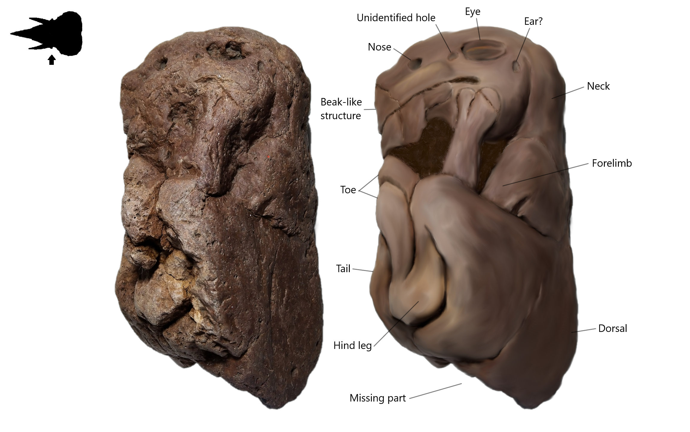
Figure 1. Lateral view (left side) on Brachylophosaurus canadensis embryo with its restoration image. The lower part of body probably gets destroyed or decayed before fully fossilized.
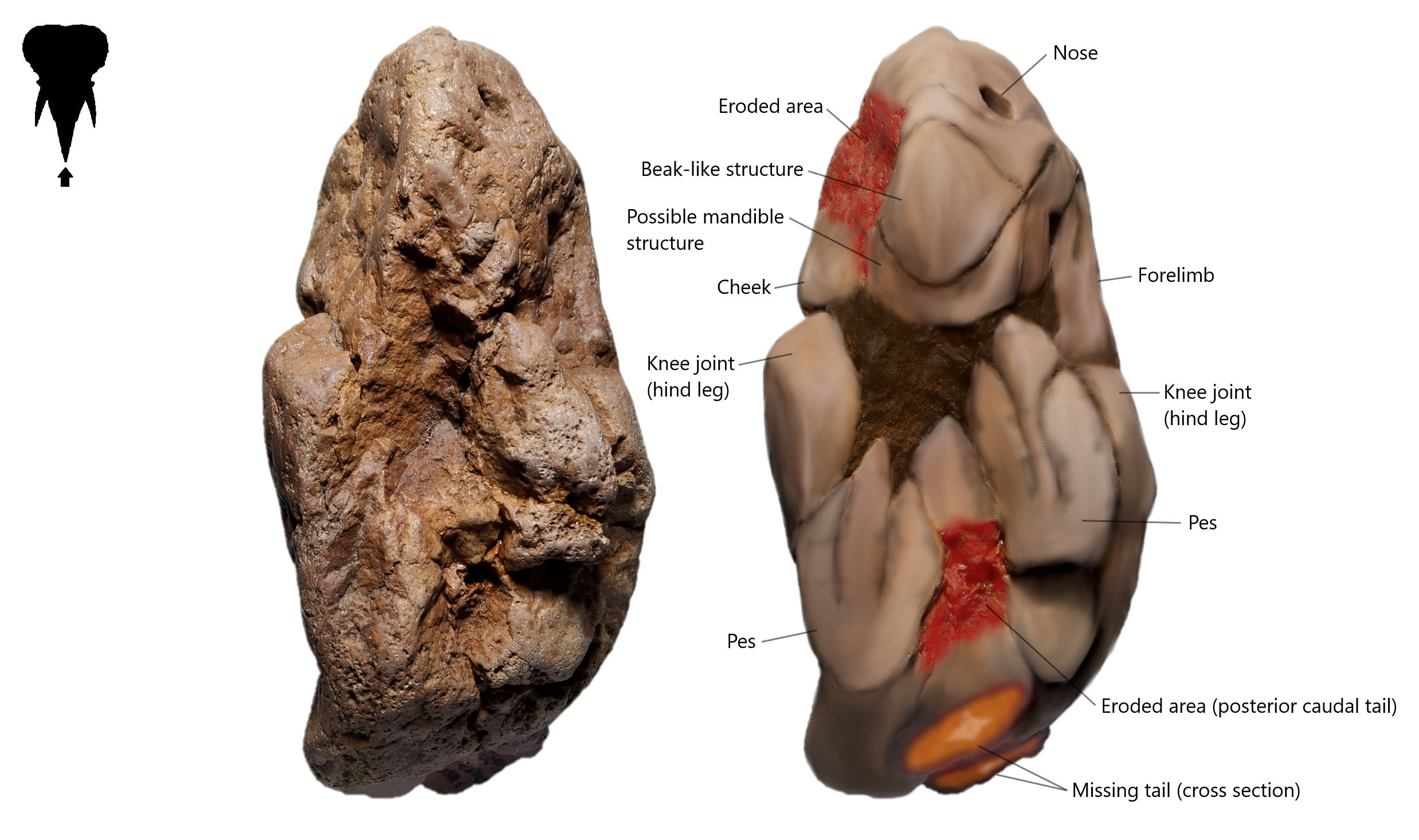
Figure 2. Front view on Brachylophosaurus canadensis embryo with its restoration image.
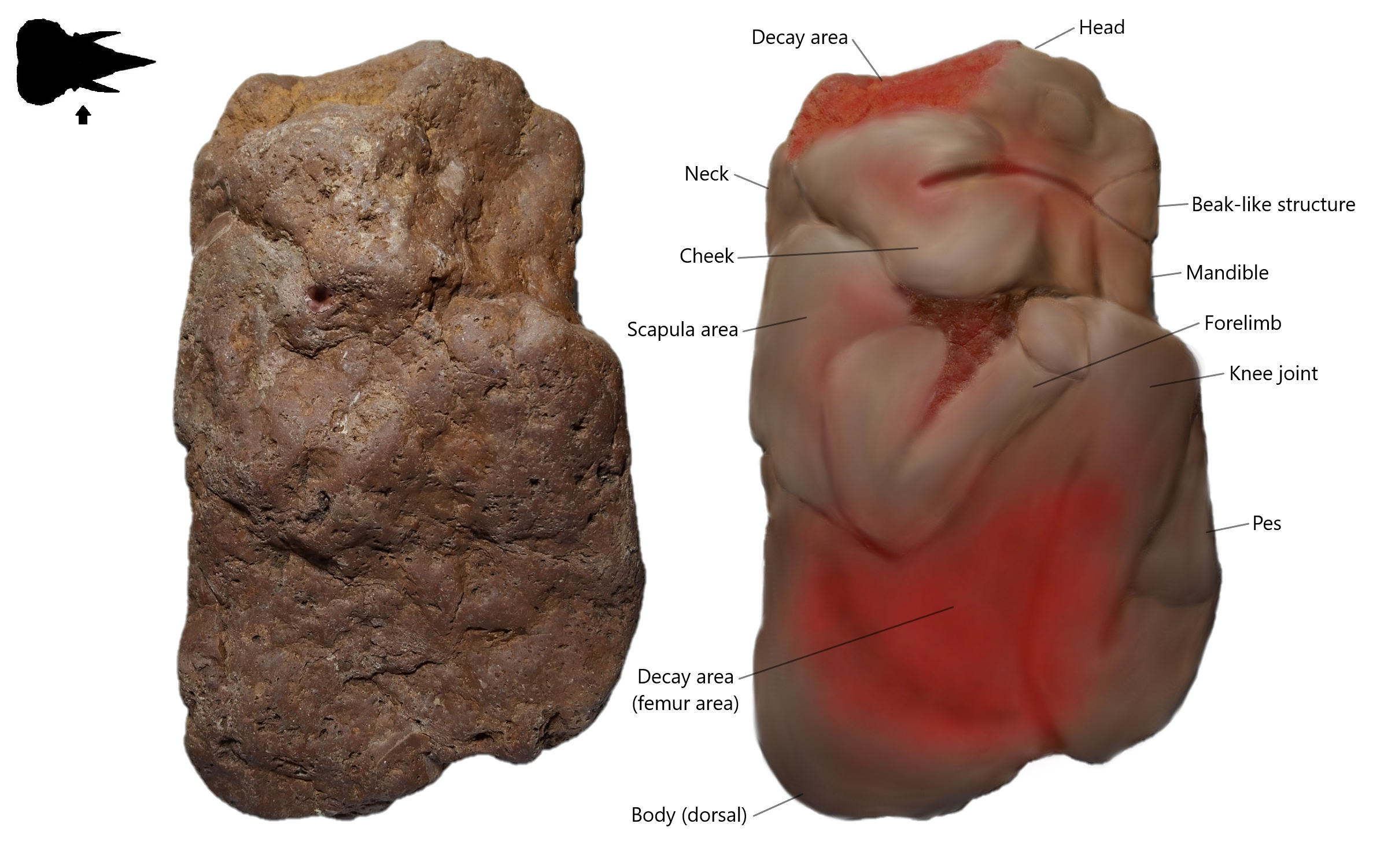
Figure 3. Lateral view (right side) on Brachylophosaurus canadensis embryo with its restoration image. Most of the surface already eroded.
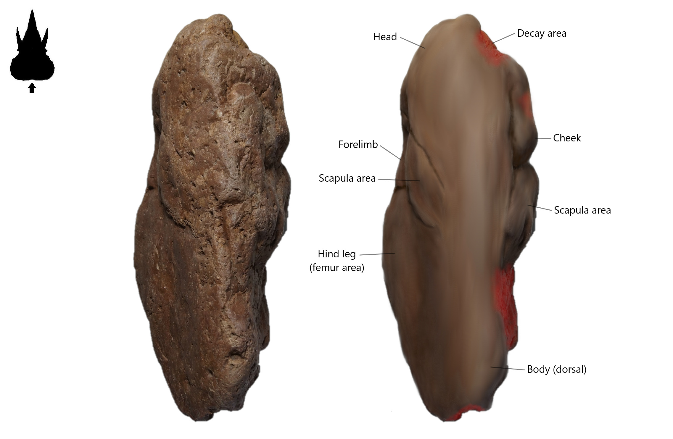
Figure 4. The dorsal view (backside of body) on Brachylophosaurus canadensis embryo with its restoration image.
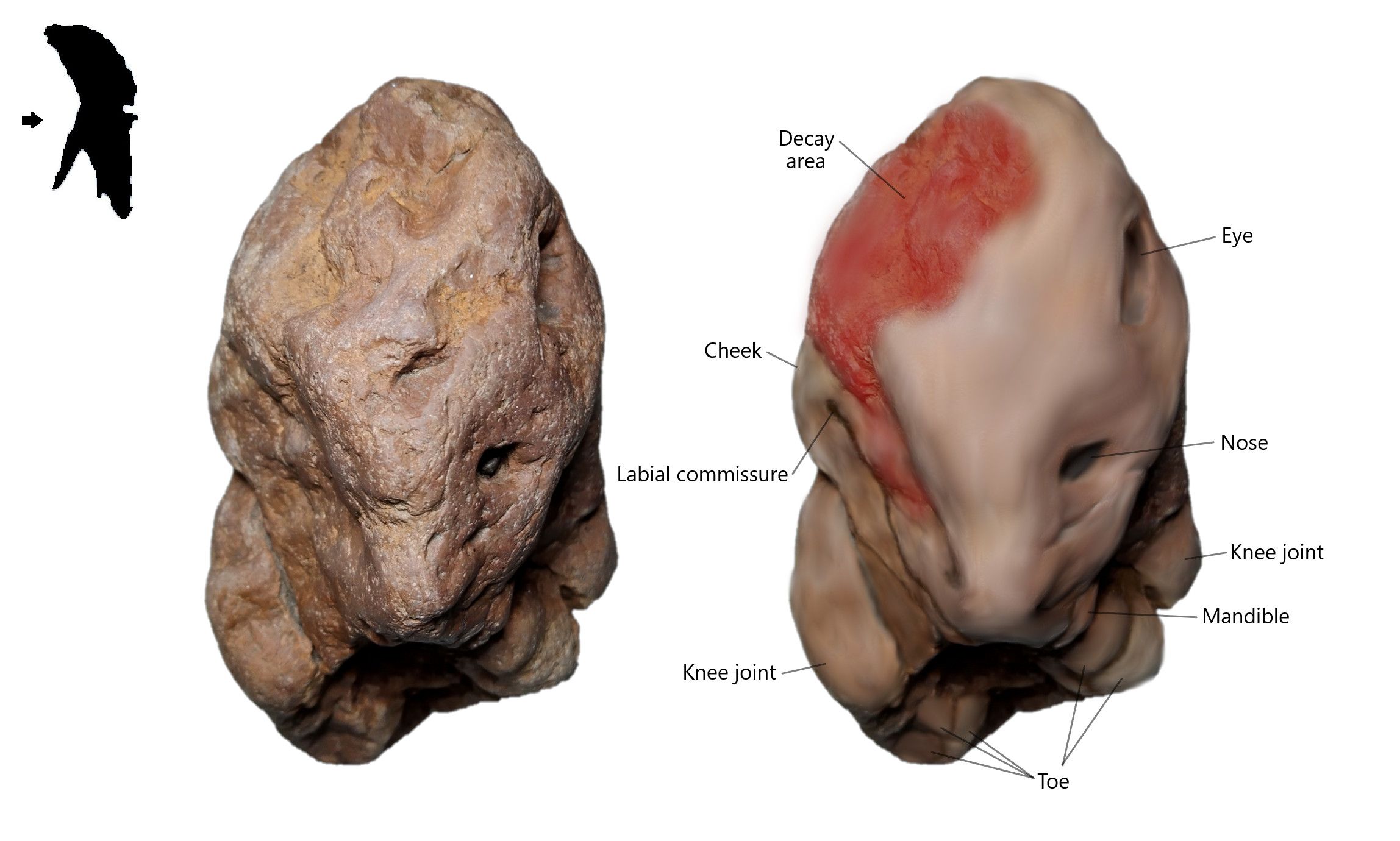
Figure 5. Top view (head) on Brachylophosaurus canadensis embryo with its restoration image.
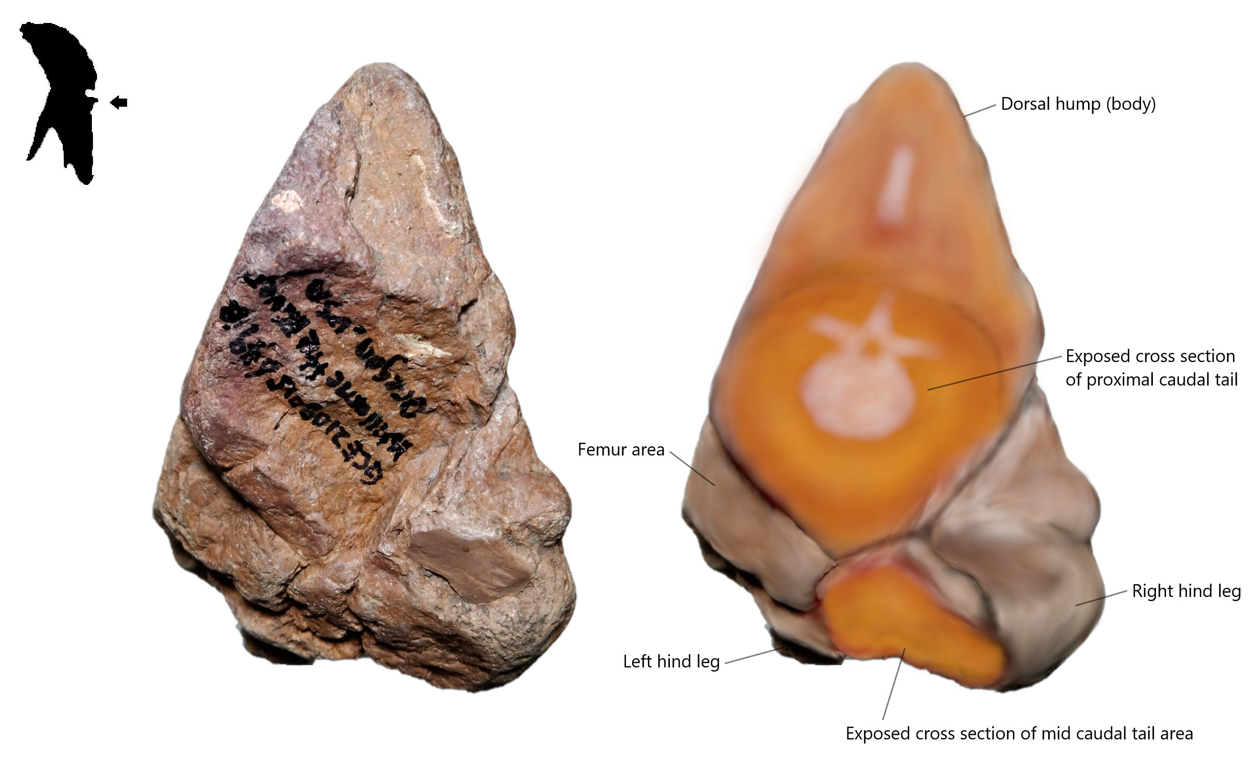
Figure 6. Bottom view on Brachylophosaurus canadensis embryo with its restoration image.
Top view of embryo head (refer Figure 5) show slightly wider than the adult fossil skull (the top view of the skull superimposed to the embryo head will not show here as limited by copyright of top skull photo) but this is typical phenomenon of the embryo. All available embryo specimens typically show wider head (means if you superimpose comparison with adult fossil skull, its eye will locate on outside of the orbit, or simply understand as eye socket) than the adult skull perhaps they start with wider head and slowly fix to adult skull shape when hatching and growing up.
Based on the shape of embryo Brachylophosaurus, the Brachylophosaurus egg is likely belong to elongated types, with estimated egg size approximate 15cm (see calculation in section Comparative Growth Stages of the embryo). The elongated types of egg shape probably more suitable for species with stiffness dorsal although the ossified tendons are yet developing in embryo stage.
The entire baby dinosaur after hatching estimate not less than 34.5 cm in length, from head to tail, according to measurement through embryo specimen without estimating its current growth stage.
Comparative Growth Stages of the embryo
Comparative growth stages, Sc is study of embryo development through comparison with common chicken embryo. The main purpose is use to determine the growth condition of fossil embryo, and use it for restoration the final state of the embryo and reconstructing the egg size. The methodology and detail already explained in volume 4, and here we will direct proceed with the calculation section to estimate the growth size of Brachylophosaurus embryo.
![Figure 7. Comparative Growth Stage Sc19 to Sc21. A reconstructed chicken embryo condition inside the egg [1].](https://petrifiedembryology.files.wordpress.com/2021/10/fig-28-19-21-note-s.jpg)
Figure 7. Comparative Growth Stage Sc19 to Sc21. A reconstructed chicken embryo condition inside the egg [1]. [**Picture contain some derivative work that still possibly copyright protected. Use with care by proper credit the source or prevent to use**]
![Table 1. A reconstructed (approximate value) Growing Percentage Chart of each Comparative Growth Stages of embryo and its yolk inside the egg based on chicken embryo [1]. Both embryo to egg size and yolk to egg size ratio (in percentage) is calculate independently based on cross section 2D form.](https://petrifiedembryology.files.wordpress.com/2021/10/table-1.png)
Table 1. A reconstructed (approximate value) Growing Percentage Chart of each Comparative Growth Stages of embryo and its yolk inside the egg based on chicken embryo [1]. Both embryo to egg size and yolk to egg size ratio (in percentage) is calculate independently based on cross section 2D form.
Based on the observation of the Brachylophosaurus embryo, the head to body size ratio appear close to adult (early stage of embryo will tend to have a bigger head than the body, and appear larger eye), tiny eye and a well-developed forelimb and hind limb all point the embryo already close to final stages and almost prepare for hatching. The embryo is estimate reach Sc19 growing stage. When the theoretical Comparative Growth Stage is finalized, we can estimate its final growing size based on few equations below.
The theoretical final growing size of Brachylophosaurus canadensis based on Sc19
= (current embryo head to tail length/Sc19 embryo percentage)*100%
= (34.5cm/90.16%)*100%
= 38.27cm
So, the theoretical hatching size of baby Brachylophosaurus approximate 38.27cm.
The size of theoretical yolk sac under Sc19
= (longest length of embryo/Sc19 embryo percentage)*Sc19 yolk percentage
= (13.5cm/90.16%)*28.25%
= 4.23cm
So, the reconstructed yolk sac size is about 4.23cm for this specimen (refer Figure 8).
The theoretical egg size of Brachylophosaurus canadensis based on Sc19
= (longest length of embryo/Sc19 embryo percentage)*100%
= (13.5cm/90.16%)*100%
= 14.97cm
So, the theoretical egg size of Brachylophosaurus canadensis is about 15cm.
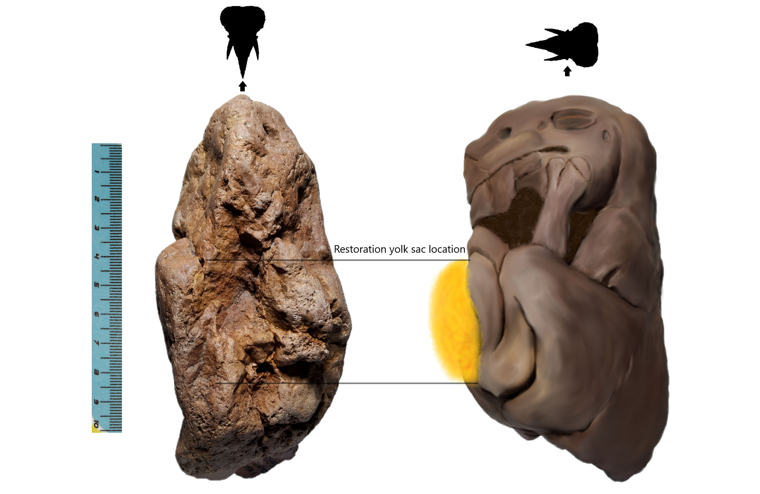
Figure 8. A reconstructed image of Brachylophosaurus embryo with its unabsorbed yolk sac locate above the abdomen. The yolk sac located area also can be roughly observed in the front view of the embryo (left image) that shows some trace of small hole. This specimen no longer preserved its yolk sac as already decayed before fully fossilized. Yolk sac contains large amount of fluid that sometimes slow the transformation into Dry Mass that has high resistance to corrosion before turn into fossil. The hypothesis of Dry Mass Fossilization please refer volume 4. (scale bar in cm)
Species Identification
Species identification of mummified specimen usually assists with the technique Superimpose Test. It using various skull comparison to identify the species. Although the actual skull of embryo within the head looks different with its adult skull, but its outer surface skin morphology will tend to have similar ratio line with its adult form. The ideology will be explaining in another chapter as it consists of various animal skull and surface comparison studies.
![Figure 9. Superimpose Test (in lateral view, left side) of restoration image with drawing of Brachylophosaurus canadensis skull [2] from Royal Tyrrell Museum.](https://petrifiedembryology.files.wordpress.com/2021/11/pev5-fig-9-note-s.jpg)
Figure 9. Superimpose Test (in lateral view, left side) of restoration image with drawing of Brachylophosaurus canadensis skull [2] from Royal Tyrrell Museum. [**Picture contain some derivative work that still possibly copyright protected. Use with care by proper credit the source or prevent to use**]
![Figure 10. Superimpose Test (in lateral view, left side) with drawing of Brachylophosaurus canadensis skull [2] from Royal Tyrrell Museum.](https://petrifiedembryology.files.wordpress.com/2021/11/pev5-fig-10-s.jpg)
Figure 10. Superimpose Test (in lateral view, left side) with drawing of Brachylophosaurus canadensis skull [2] from Royal Tyrrell Museum. [**Picture contain some derivative work that still possibly copyright protected. Use with care by proper credit the source or prevent to use**]
Figure 10 shows a Superimpose Test of specimen GCE2108015649B with Brachylophosaurus canadensis skull from Royal Tyrrell Museum. Most preserved trace lines show identical with the skull. One of the distinct morphology between crested and non-crested Hadrosaur is non-crested Hadrosaurid poses a very large nose hole. Although it sounds weird, but Brachylophosaurus show its labial commissure opening until below the eye socket (orbit). I have examined few Hadrosaurid mummified specimens most of them show some traces of labial commissure opening extend until the lower position of eye socket. The observation shows a different result than our general understanding of Hadrosaur mouth morphology based on existing mammals, it could be an extremely rare occurrence in animals but it still possible to occur.
The lower skin extension of non-crested Hadrosaur
One of the new findings about the non-crested Hadrosaurid is some of their species pose a very large skin extended area below the mandible (refer Figure 11). The skin extended area usually 2x the size of mandible and typically show on species having a curve nasal (nose area). Please refer my outcoming book for other specimens. The snout area of Hadrosaurid poses some small skin hump (possible flexible and leathery like a bill of Platypus) which follow on some specific skull area and extend until a beak-like structure below the premaxilla area that sometimes refer as rhamphotheca. When closely looking on the head morphology of Hadrosaurid, which pose a beak-like structure like a parrotfish, and having a very large skin extended area below the mandible, non-crested Hadrosaurid are likely feeding on vegetation growth under the water, and they might contain a salt gland (refer figure 9) to secrete the extra salt out from the body. But this hypothesis is still under study.
![Figure 11. The head morphology of Brachylophosaurus canadensis [2] in lateral view (left). The restoration structure based on specimen GCE2108015649B refer as embryo Brachylophosaurus.](https://petrifiedembryology.files.wordpress.com/2021/11/pev5-fig-11-note-s.jpg)
Figure 11. The head morphology of Brachylophosaurus canadensis [2] in lateral view (left). The restoration structure based on specimen GCE2108015649B refer as embryo Brachylophosaurus. [**Picture contain some derivative work that still possibly copyright protected. Use with care by proper credit the source or prevent to use**]
The beak-like structure of Hadrosaurid
The beak of Hadrosaurid sometimes refer as rhamphotheca and typically they trust made up from keratin like the beak of existing birds. But close view on the skull of Hadrosaurid, the beak-like structure of Hadrosaurid might not really made up from keratin. Most keratin types beak are built up from a bony base which surface will surrounded with a lot of grooves, like example below at Figure 12A, the beak structure from Gastornis giganteus, and the beak structure of Triceratops prorsus from Figure 12B. When looking at the mouth structure of Brachylophosaurus canadensis in Figure 12C, it did not show an obvious groove structure and a bony base for keratin attachment. Instead, they probably build up from a fused enamel-like tooth like the beak-like tooth from parrotfish (refer Figure 16) that extends from the premaxilla. Although they might not build from the same material (fluorapatite) like a parrotfish, but could be a close similar structure of low strength material than teeth.
![Figure 12. Lateral view (left side) on snout structure of (A) flightless bird Gastornis giganteus [3]; (B) Triceratops prorsus [4]; and (C) Brachylophosaurus canadensis [2].](https://petrifiedembryology.files.wordpress.com/2021/11/pev5-fig-12-s.jpg)
Figure 12. Lateral view (left side) on snout structure of (A) flightless bird Gastornis giganteus [3]; (B) Triceratops prorsus [4]; and (C) Brachylophosaurus canadensis [2]. [**Picture contain some derivative work that still possibly copyright protected. Use with care by proper credit the source or prevent to use**]
![Figure 13. Front view on the beak-like structure of Edmontosaurus mummy SMF R 4036 [5].](https://petrifiedembryology.files.wordpress.com/2021/11/pev5-fig-13-s.jpg)
Figure 13. Front view on the beak-like structure of Edmontosaurus mummy SMF R 4036 [5]. [**Picture contain some derivative work that still possibly copyright protected. Use with care by proper credit the source or prevent to use**]
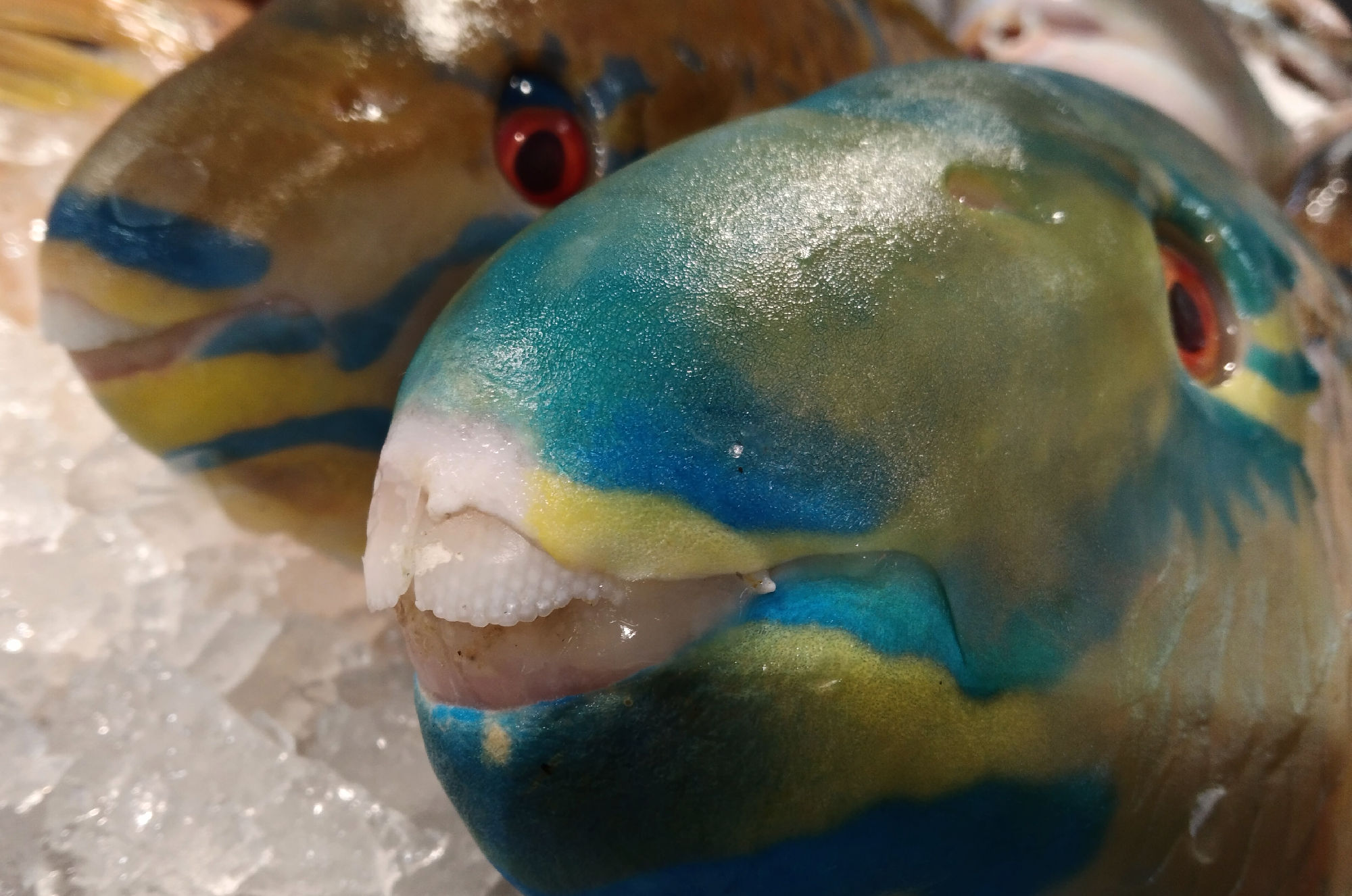
Figure 14. Front view on the beak-like teeth of parrotfish.
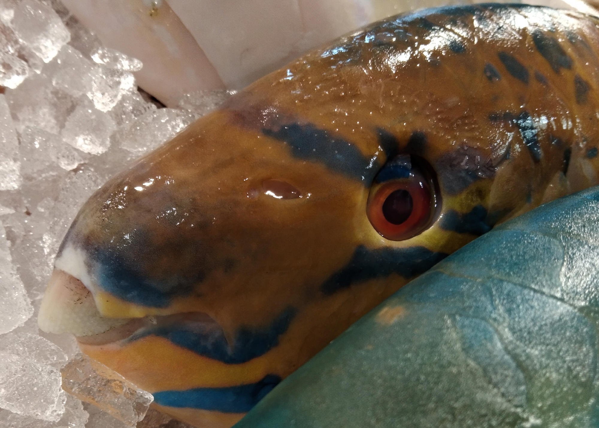
Figure 15. Lateral view (left side) on the beak-like teeth of parrotfish.
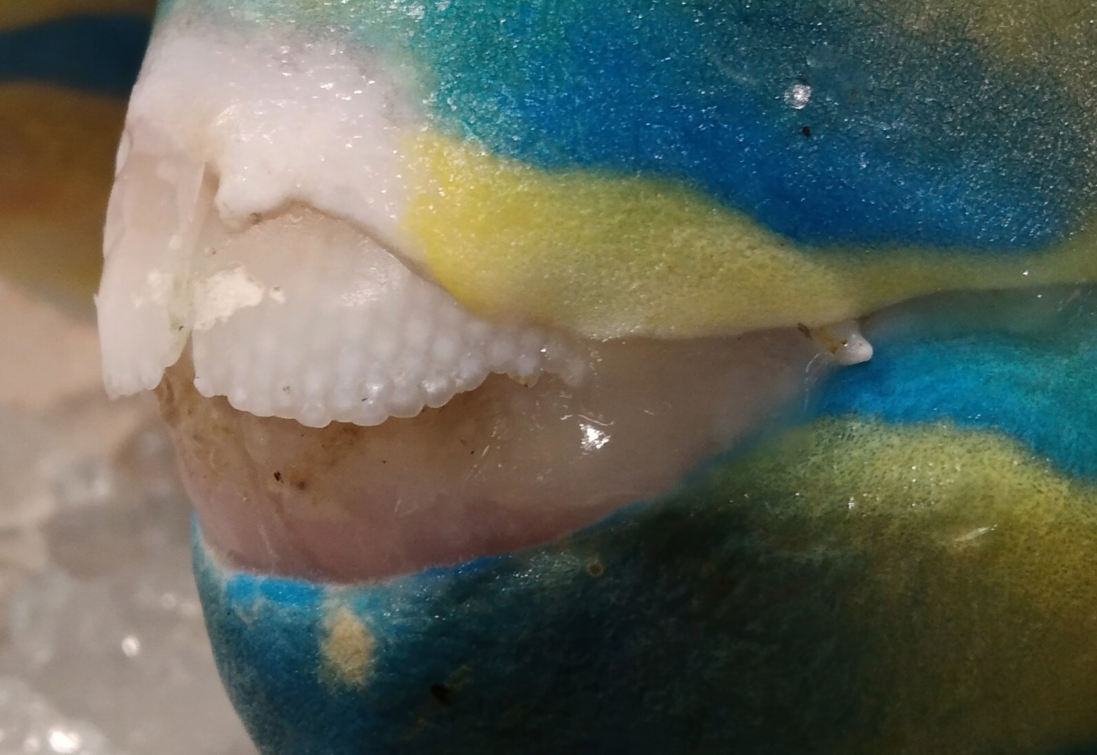
Figure 16. Close-up view on the beak-like teeth structure of parrotfish.
Why dinosaur embryo always appear detail only in one side?
If you ever searching and collect the Agatized dinosaur embryo, you will probably notice that the embryo always appears a detailed structure only on one side, the opposite side of embryo will tend to show a fuzzy texture (Figure 17). But this is normal phenomenon of Agatized embryo when they are formed from Dry Mass Fossilization (refer volume 4 for the theory).
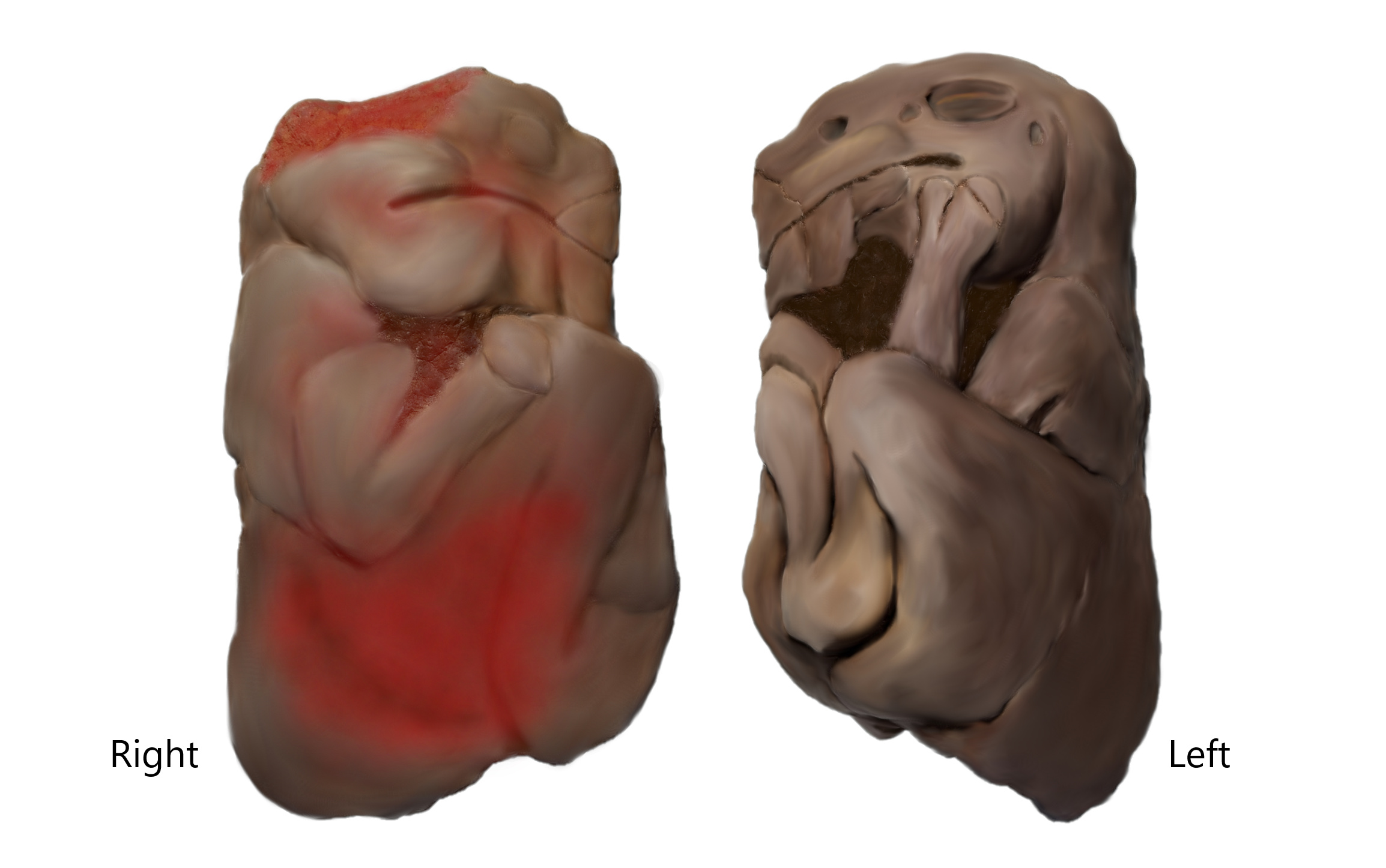
Figure 17. The lateral view of restoration image on Brachylophosaurus canadensis embryo in right and left.
To understand this, please refer Figure 18 that shows a general structure of most embryo lay on top of albumin or yolk sac within the egg. Most of the eggs will lay horizontally on the ground. Under initial stage, the overall fluid distributed evenly within the egg. When the embryo dead in the raining or dry season, the growth activity within the egg stop. The remaining fluid level within the egg will slowly evaporate and cause the formation of dry mass embryo which has high resistance from decay. But due to different level (Figure 19) within the egg exerting different rates to dry mass formation, the top level of egg typically has the higher speed of dry mass formation, due to low fluid composition. The lowest the total fluid level, the higher the rate of dry mass formation; the higher the fluid level, the lowest the rate of dry mass formation. When you found a large portion of embryo body is destroyed, they probably cause by early presence of cracking hole on the eggshell that protect the internal content, which lead to a large portion of erosion and decay on the cracking hole area (Figure 20). Shrinking and becoming flat on the bottom side is also one of the common phenomena caused by gravity effect on decay area and cause the surface detail completely turn blank and flat.
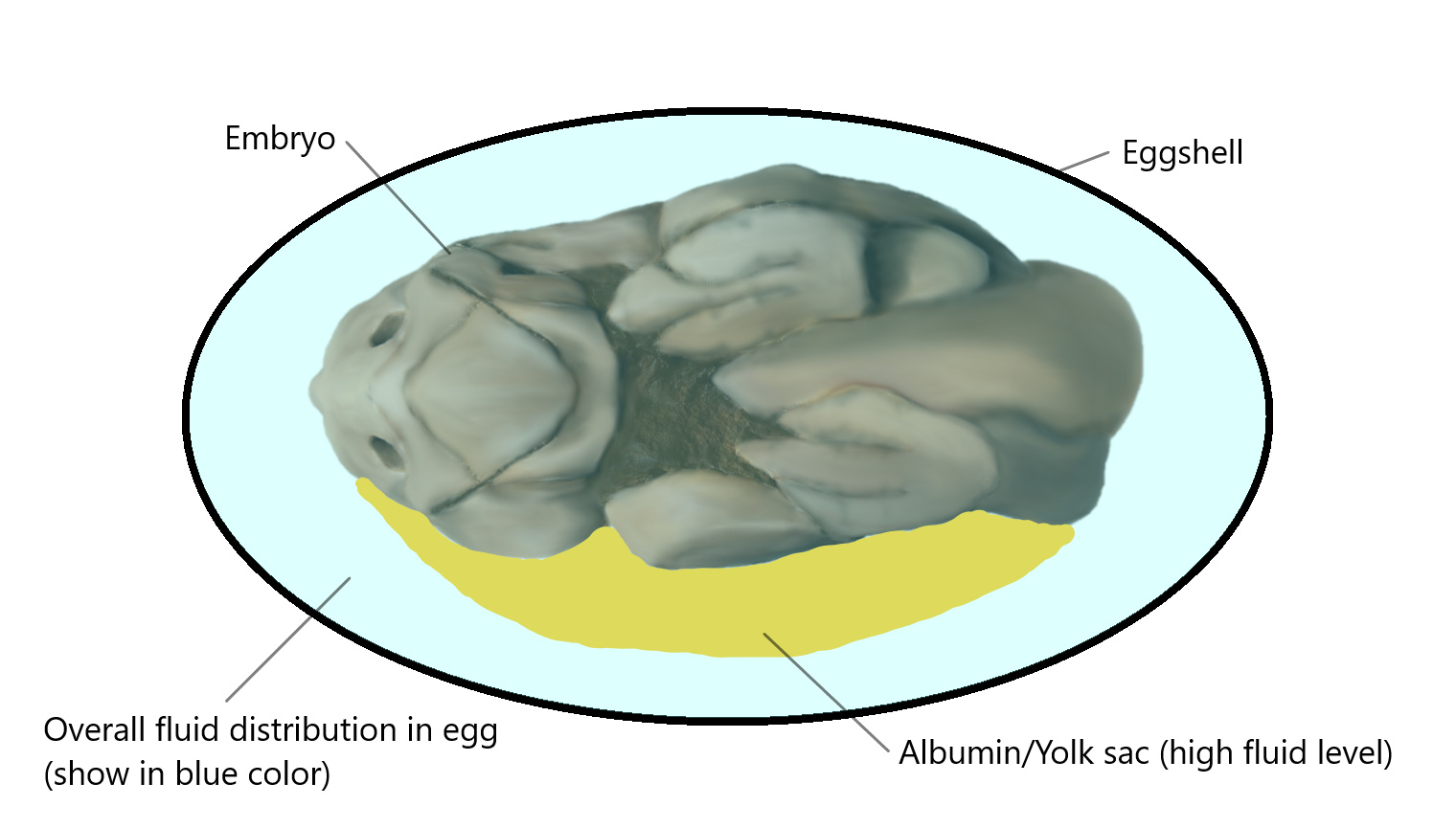
Figure 18. The general structure of dinosaur embryo within egg (in front view). The blue color shows the overall distribution of fluid within egg in initial stage before the embryo dies within the egg.
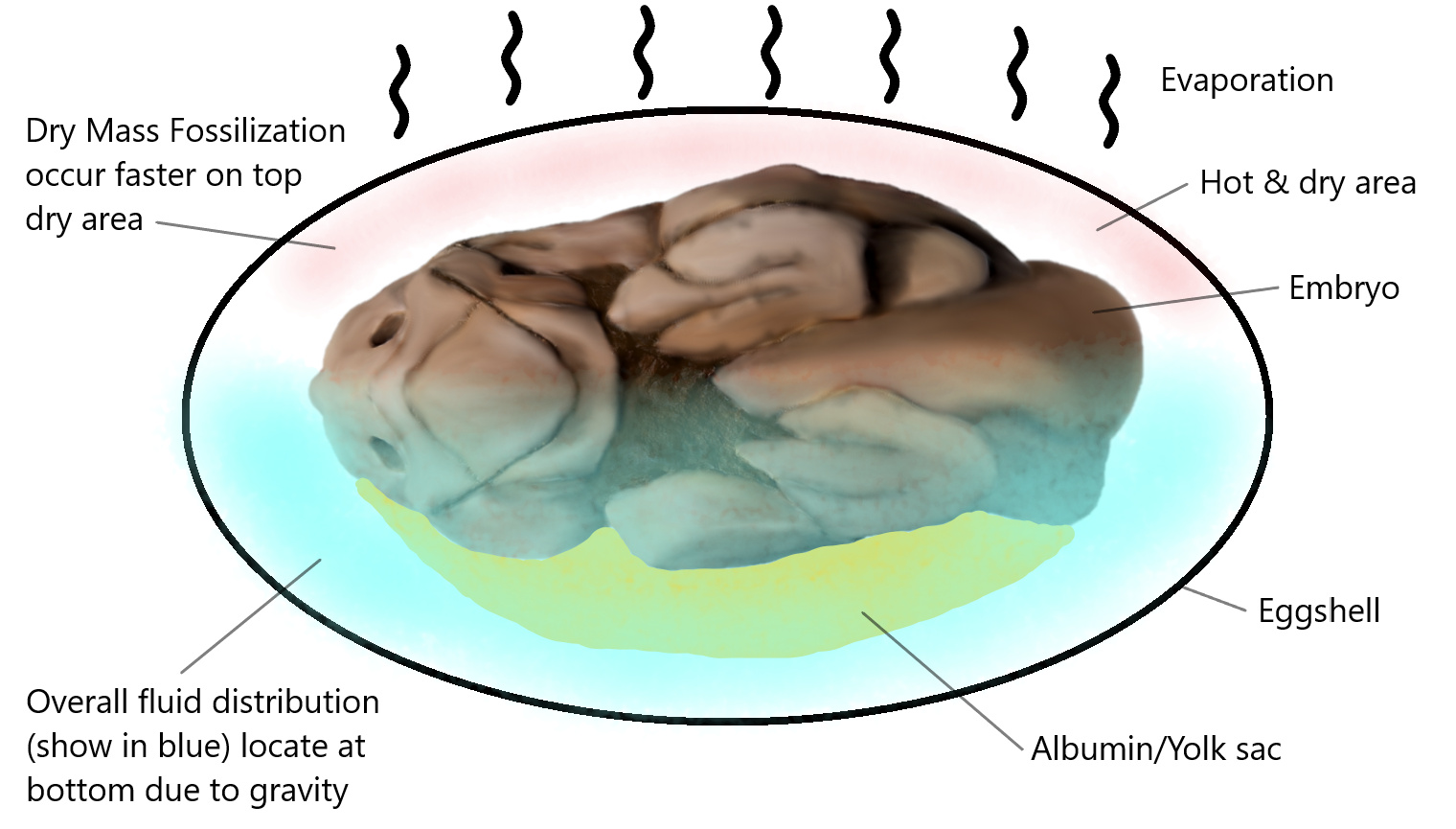
Figure 19. The picture shows how the total fluid distribution within the egg when the embryo dies in dry season. Most fluid will locate at bottom level due to gravity pull which has the slowest rate to form dry body mass that leads to high decay rate.
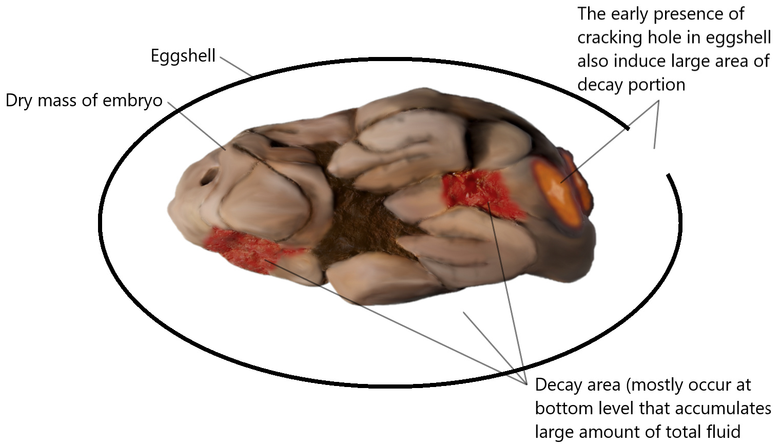
Figure 20. Most decay area will occur at the bottom part that accumulates large amount of fluid with lowest rate forming dry body mass. The early presence of cracking hole in egg will also cause large area of body part to decay.
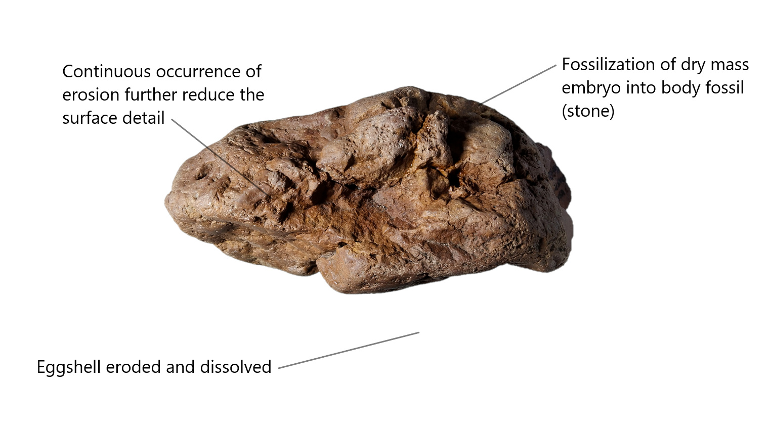
Figure 21. Fossilization of dry mass embryo and turn into ordinary rock. The continuous natural erosion causes a further reduction in surface clarity. Most eggshell will erode but if eggshell is preserved, they will usually form a thin film-like layer on top of the embryo with distinct color.
The shrinking effect will also occur during the formation of dry body mass due to loss of body fluid, the surface becomes dry and shrinking. The fully develop the embryo, the lower the shrinking effect due to the body of full developed embryo contain more protein, muscle and flesh and less water composition than the early-stage embryo that contain higher composition of water that will shrink more when water evaporates.
Declaration
As researcher, I’m hereby to declare the specimen is fully in nature form without perform any artificial modification or as man-made stone crafts other than perform a normal cleaning and repair on a small crack on hind limb section.

The roughly size of embryo compares to my hand as reference without any editing on image.
Acknowledgments
Special Thanks to one of my friend, Matthew G. Tuma, from United States, which provide some good specimens for study and improve my understanding on some missing link of Hadrosaur.
More books available at https://petrifiedembryology.wordpress.com, a nonprofit open science study of mummify dinosaur specimen.
References
- CheeEng Goo. Petrified Embryology Volume 4: The Frozen Baby Dinosaurs – Procheneosaurus praeceps. Smashwords. 4:P20 (2021)
https://petrifiedembryology.wordpress.com/petrified-embryology-volume-4-the-frozen-baby-dinosaurs-procheneosaurus-praeceps/ - Etemenanki3. Brachylophosaurus canadensis skull (original). From the Oldman Formation, Milk River, Alberta. On display at the Royal Tyrrell Museum, Alberta, Canada. Wikimedia Commons. (2017)
https://commons.wikimedia.org/wiki/File:Brachylophosaurus_skull_Royal_Tyrrell.jpg - Vince Smith. Skeleton of Gastornis giganteus, a large flightless bird from the Eocene found in Wyoming. Wikimedia Commons. (2011)
https://commons.wikimedia.org/wiki/File:Gastornis,_a_large_flightless_bird_from_the_Eocene_of_Wyoming.jpg - Daderot. Triceratops prorsus, Carnegie Museum of Natural History, Pittsburgh, Pennsylvania, USA. Wikimedia Commons. (2009)
https://commons.wikimedia.org/wiki/File:Triceratops_prorsus_-_IMG_0697.jpg - Etemenanki3. The Edmontosaurus mummy SMF R 4036 on display at the Senckenberg Museum, Frankfurt, Germany. Wikimedia Commons. (2013)
https://commons.wikimedia.org/wiki/File:Senckenberg_mummy_6.jpg


More issues available on home page


